If you buy learning content for a nursing school, an allied-health programme, a fitness or first-aid training business, a medical-device company's sales team, or a hospital's patient-education service, you are paying for one thing: people understanding the body well enough to act. Most of the material that does that job is still flat — textbook plates, slide decks, a plastic model on a shelf, a leaflet handed to a patient before a procedure.
Flat material has costs that do not show up on the invoice. Learners struggle to build a 3D picture from 2D drawings, so training takes longer. Licensed 3D anatomy tools exist, but they are often seat-licensed, installed software that is awkward to put in front of a patient or a sales prospect. And popular "facts" — the famous 100,000 km of blood vessels, alveoli the size of a tennis court — circulate in course material long after anyone has checked them, which is a credibility risk for any organisation that teaches health.
What a digital twin means here
A digital twin in the full sense mirrors a specific, live system. Our Human Body Explorer is not a twin of your body or a patient's body. It is a digital model — generation 1 in our framework: a faithful 3D structure from authoritative, measured data, with facts that are known and fixed and no live sensor state.
For education and explanation, that is the right level. The value is accuracy, traceability and the ability to explore and ask. A buyer would need more — a patient-specific model from their own imaging, or physiology that responds to real measurements — only for clinical planning or monitoring, which is a regulated use and a different product altogether.
What we built
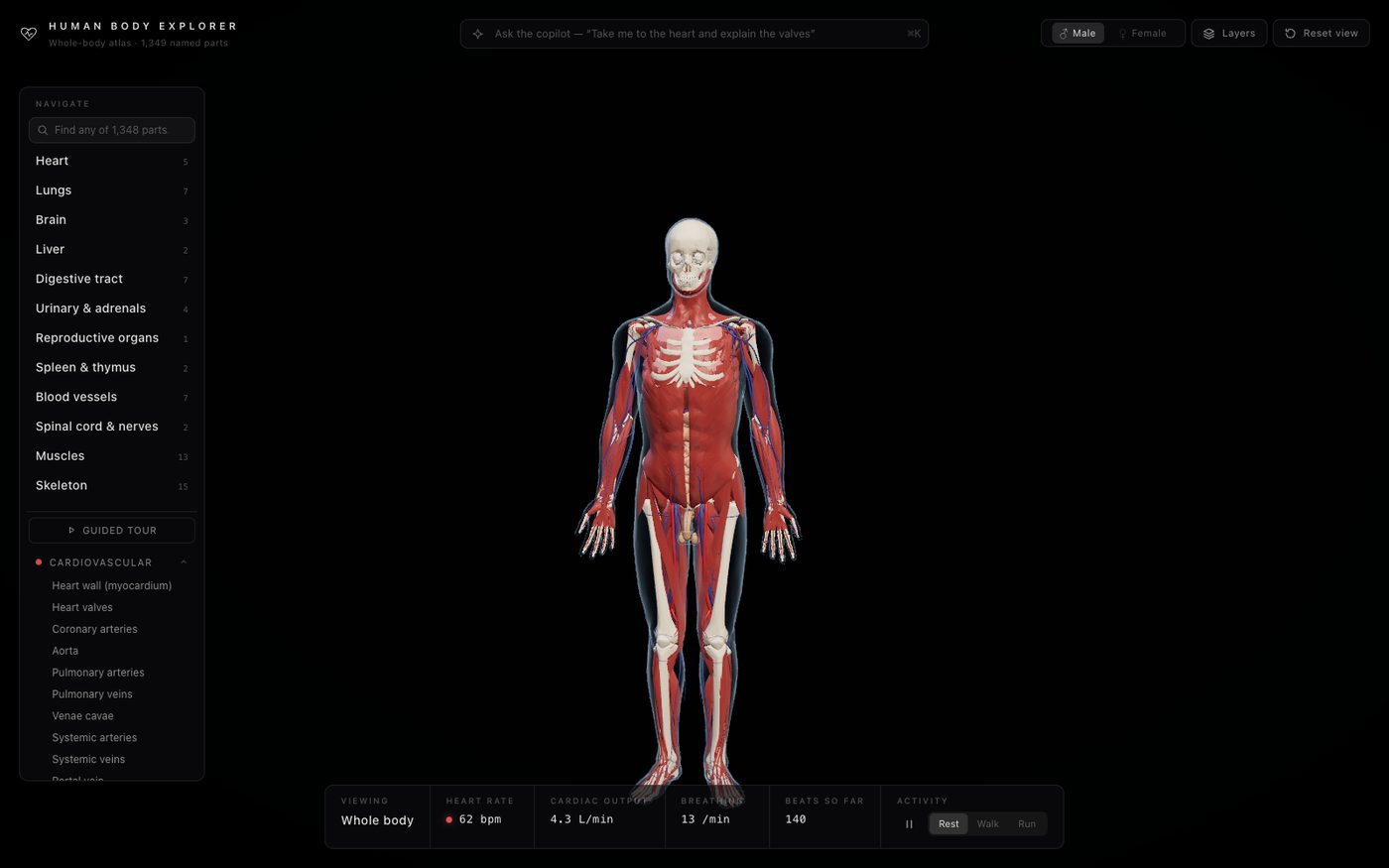
Human Body Explorer is an interactive 3D atlas of the whole body that runs in the browser. It contains 75 structures across ten body systems — cardiovascular, respiratory, nervous, digestive, urinary and endocrine, lymphatic, muscular, skeletal, reproductive and integumentary — subdivided into about 1,300 individually named parts you can hover, click and search: the heart by chamber and valve, the brain by lobe and gyrus, each rib and vertebra, the liver by segment, roughly 200 named arteries and as many veins, every named muscle.
- Measured geometry. The meshes come from BodyParts3D, surfaces segmented from a whole-body MRI of one adult volunteer, each carrying its standard anatomical (FMA) term. A female module from the HuBMAP Human Reference Atlas can be switched in.
- Audited facts. Every structure has a card with function, size, weight and a short description. The cards were audited in September 2026 against clinical and textbook references, with ranges where sources differ, and popular but unmeasured figures were removed or corrected.
- A copilot. Ask in any language — "take me to the heart and explain how the valves work", "show me the skeleton without the skin", "肝脏是做什么的?" — and Claude on Amazon Bedrock opens the right section and flies the camera there while it explains, under the same factual rules as the cards. A built-in guide answers when the service is offline.
- Teaching tools. A guided tour of the major organs; dissection-style focus that fades whatever is in the way; skin opacity and a cross-section slider through the chest; a beating heart and breathing lungs with rest, walk and run levels and illustrative vitals.
Questions it helps a buyer answer
- Can learners explore anatomy in 3D on the devices they already have, without installing software?
- Can we show a patient, before a procedure, exactly which structure we are talking about — and let them ask their own question in their own language?
- Can we stand behind every figure we teach, and show where it came from?
- Can one tool serve classroom teaching, self-study and front-line explanation?
What changes
Before: flat diagrams, plastic models and slide decks; facts copied from course to course; 3D tools locked to a few licensed seats; explanations that depend on who happens to be explaining.
After: a single link opens a measured, searchable 3D body on a laptop, tablet or phone. Learners move from "look at the picture" to "go and find it". Patient conversations start from the actual structure. Facts have been checked and the checking is documented, so the content is something an educator can defend.
Getting there
For a training or patient-education edition we would want to know:
- your audience (students, staff, patients, customers) and what they must understand afterwards;
- the body regions, procedures or products to focus on;
- your languages, accessibility requirements and devices;
- your review process for clinical content, and how the AI guide should be constrained for your setting.
As with every engagement: discovery to agree audiences and learning outcomes; a prototype with curated tours for your topics; then a production edition with your content review, your branding and the guardrails your clinical or education governance requires.
Questions for any anatomy-content vendor: Where does the geometry come from, and is it measured or sculpted? Who checked the facts, against what, and when? What does the AI do when it is unsure or asked for medical advice? Can it run without the AI service?
Honest limits
This is an educational model, not a diagnostic or clinical tool, and nothing in it is medical advice. The body is a single adult male scan; the female organs come from a different reference atlas, so they are correct in shape and placement but not from the same body, and one structure for which no open imaging-derived model exists is shown as a labelled schematic. The heartbeat and breathing animation uses textbook values and is illustrative, not a physiological simulation. The copilot is an AI and should sit inside your own clinical-content review.
See it
- Live: body.octopustwin.com
- Case study: Human Body Explorer
- Talk to us about a training or patient-education edition: contact